Are you are seeing your ophthalmologist or optometrist regularly for your eye health? If you do, you experience some of the most exciting technological instruments in medicine.
The auto-refractor is often the first instrument used to test your eyes. It assists the doctor in approximating what you need for an eyeglass prescription by bouncing light off the retina and examining the reflections. Because "refraction" for updated eyeglasses often costs money out of pocket, patients often decline it being done. Unless you're thrilled with your vision, I recommend you do this at least every year, and sometimes more frequently.
The Slit Lamp exam is the biological microscope that allows the eye doctor to examine your eyes in 3D, with very high magnification. He or she uses the slit lamp to assess for dry eye syndrome, corneal health, cataracts or the state of your lens implants if you've had cataract surgery and more. One of the most popular slit lamps is the Haag-Streit, a Swiss-made wonder that has remained relatively unchanged in design for many years.

Retinal cameras now use digital imaging techniques, and many can even take photographs through undilated pupils, a real convenience for patients. Images of the tiny macular area can be enlarged on the computer screen, showing the doctor areas of drusen, bleeding, atrophy or other abnormal findings. Macular Degeneration is readily seen with retinal cameras, and it allows the doctor to follow your progress more accurately over time.

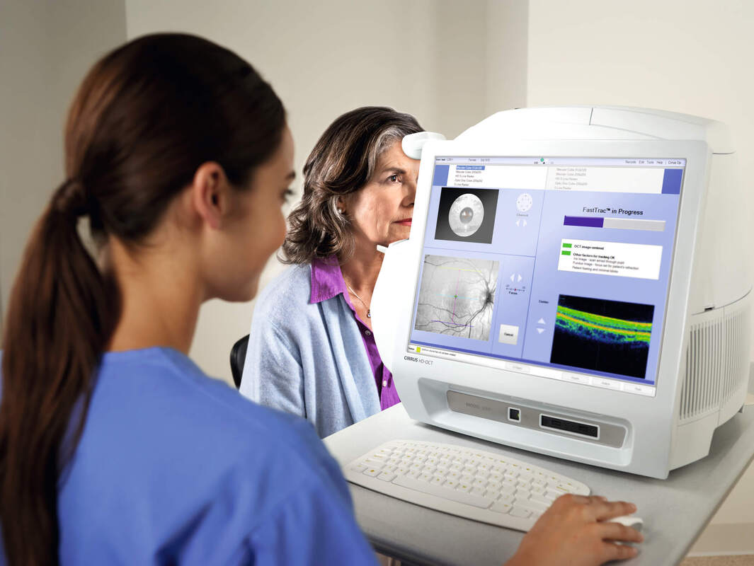
Finally the OCT, or Optical Coherence Tomographer, such as the one below from Zeiss, has changed the way that macular degeneration is assessed. Without having to inject dye into the patient's eye, OCT performs imaging by measuring the echo time delay of reflected light. It is able to then examine the layers of the retina, and most importantly to asses whether the macular degeneration is the dry or wet version.