Goniscopy is a critical element to any glaucoma exam. Dr. Paul Krawitz explains why.
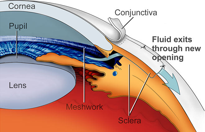
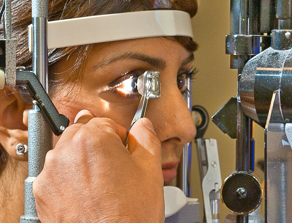
The placement of a gonioscopy mirrored lens on the surface of the cornea allows the eye doctor to examine an important structure that cannot be seen by penlight or even the slit lamp biomicroscope. The structure is called the anterior chamber angle, and it contains the peripheral iris, ciliary body, trabecular meshwork and peripheral internal cornea.

In patients with specific eye findings, including narrow anterior chamber, elevated eye pressure, ocular hypertension, glaucoma, pigmentary dispersion syndrome, pseudoexfoliation syndrome, uveitis, eye trauma, eye tumors, proliferative diabetic retinopathy, central retinal vein occlusion and more, gonioscopic examination is a critical element to a thorough evaluation of eye health and proper diagnosis.
Alarmingly, studies in the scientific literature state that nearly 50% of community ophthalmologists fail to perform this important examination, due to technical inability or disinterest.
Perhaps the most important function of gonioscopy is in determining whether a patient's elevated eye pressure is due to an "open angle" or "closed angle" mechanism. In the Western hemisphere, most glaucoma and elevated pressure is due to an open angle mechanism, which can include primary open angle glaucoma, ocular hypertension, pseudoexfoliation syndrome and pigmentary dispersion syndrome. In the East, however, a large percentage of patients with elevated eye pressure - perhaps as high as 50% - are due to a closed or narrow angle mechanism.
The initial treatment for elevated eye pressures associated with an open angle mechanism include monitoring, eyedrops, selective laser trabeculoplasty, and less commonly surgery, while the treatments for narrow angle and elevated eye pressure due to narrow angle include laser iridotomy and less commonly, laser iridoplasty.
Patients with narrow angles who have been treated incorrectly for open angle glaucoma using eyedrops may have their pressure controlled for several years. However, with time, the iris and cornea can "stick together," a condition known as peripheral anterior synecchiea. This effectively seals closed the trabecular meshwork drain of the eye permanently, and may be associated with severe and permanent vision loss and the need for major eye surgery.
Patients with a family history of glaucoma, narrow anterior chamber angles or elevated eye pressures are urged to insist of their doctors that a gonioscopic examination be performed. Laser iridotomy can effectively prevent most narrow angle patients' glaucoma from progressing.