
WHICH AREDS 2 IS RIGHT FOR YOU?
NATURALLY-SOURCED EYE VITAMINS SINCE 2001
VisiVite Eye Vitamin Formulas are based on proven research in the most respected medical publications, including:
- The National Eye Institute's Age-Related Eye Disease Study (AREDS)
- Age-Related Eye Disease Study 2 (AREDS2)
- The Lutein Antioxidant Supplement Trial (LAST)
FREE SHIPPING
FOR ORDERS $50 AND UP
Real and authentic reviews
HEAR FROM VISIVITE CUSTOMERS
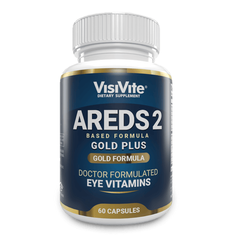
AREDS 2 Gold Plus
l know for a fact my eyes are doing great because of VisiVite
Glenn Miller
AREDS Lutein+
Been using for 15 + years and has kept my eyes in tip top shape for a 70+ year old!!
Walter Mansolillo
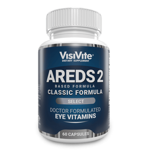
AREDS 2 Classic
VisiVite has helped me maintain the excellent vision I achieved after cataract surgery 10 years ago.
Stanley Cohen
TOP SELLERS
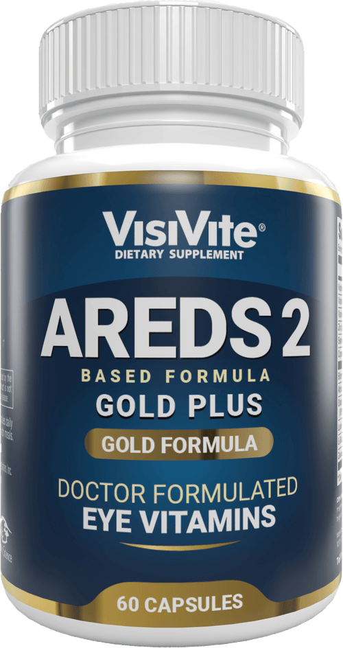
AREDS 2 PLUS+ Gold Eye Vitamin
The top tier gold standard. Contains 50% more Lutein and 138% more Zeaxanthin.
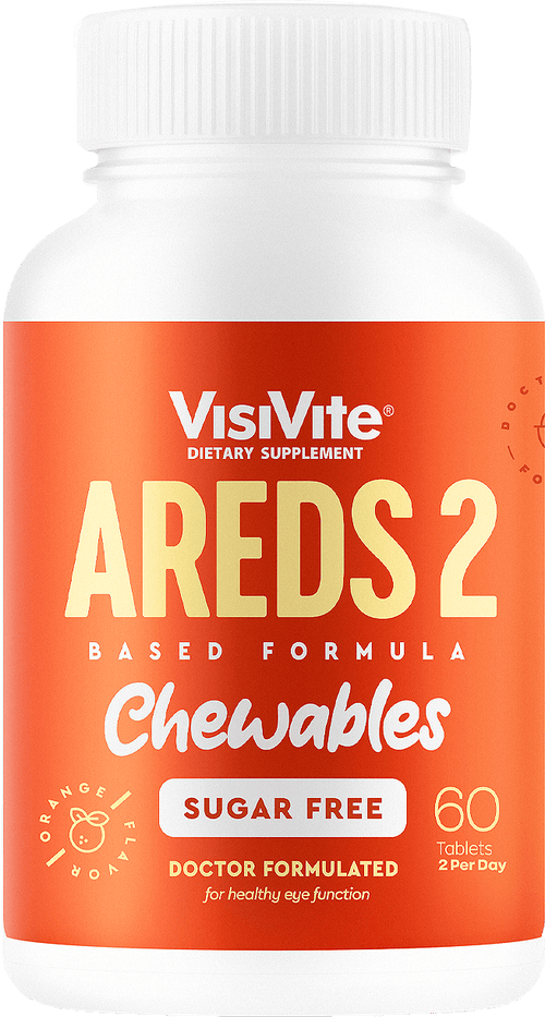
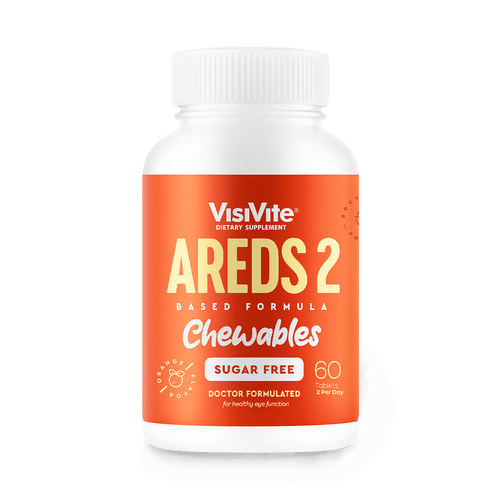
Sugar*Free AREDS 2 Chewable
The tasty AREDS 2 you'll look forward to taking every day.
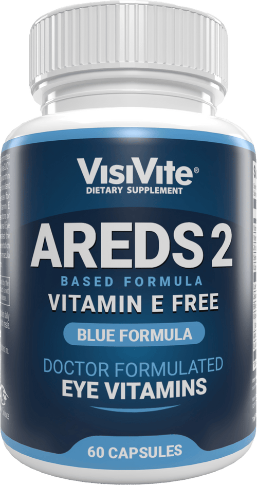
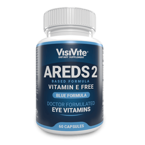
AREDS 2 E-Free Blue Eye Vitamin
AREDS 2 without Vitamin E. Made for those taking anticoagulants.
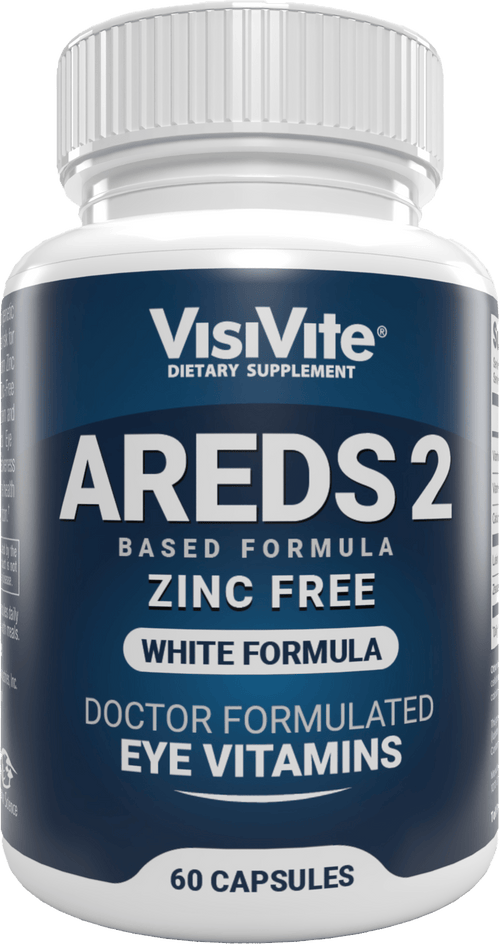
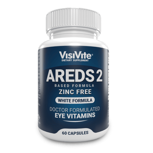
AREDS 2 Zinc-Free Eye
Designed for those with Zinc sensitivities. The AREDS 2 ingredients without Zinc.
why visivite?
A drugstore store clerk can't advise you on AMD or dry eye syndrome. A "Big Box" discount store doesn't know the difference between natural and synthetic vitamin E. And ordinary vitamin manufacturers, juggling thousands of different products,
simply can't match Vitamin Science's
specialization in eye vitamins.
DOCTOR FORMULATED
VisiVite Eye Vitamin VisiVite was founded by Dr. Paul Krawitz, a world-famous eye doctor who originated multiple trademarks and patents for innovative eye supplements. Dr. Krawitz spent over 20 years carefully researching and creating the original formulas for the 18 eye vitamin products that have been the foundation for excellence in vision support.